|
University of Ioannina, Greece |
|||||
|
|
|
||||
|
ED-XRF Spectroscopy Unit |
|||||
|
|
|||||
|
|
|||||
|
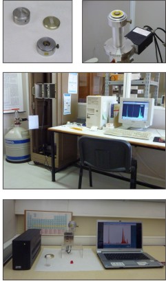
EQUIPMENT |
|||||
|
In the XRF technique, the analyzed sample is irradiated with primary X-rays and subsequently emits fluorescent X-rays, which are characteristic of the atoms present in the sample and are used for the elemental analysis. Thus, any XRF spectroscopy equipment typically consists of an X-ray source for sample excitation and a detection system for detecting the secondary fluorescent X-rays. The Unit maintains two home-built ED-XRF spectroscopy arrangements, with the following features: |
|||||
|
Exciting radiation: |
|
||||
|
|||||
|
Detectors: |
|||||
|
|||||
|
|||||
|
Software: |
|||||
|
|||||
|
Elemental range: |
|||||
|
|||||
|
Sample preparation facilities: |
|||||
|
Standard laboratory equipment - grinders, mortars, sieves, furnace, balance, pellet press, freeze drier - is used for sample handling and preparation, while chemical reagents and standard reference materials for quantitative analysis are also available. |
|||||
|
|
|||||
|
| About us | Equipment | Services | Research activities | Links | |
|||||